Technology to drive breakthrough science
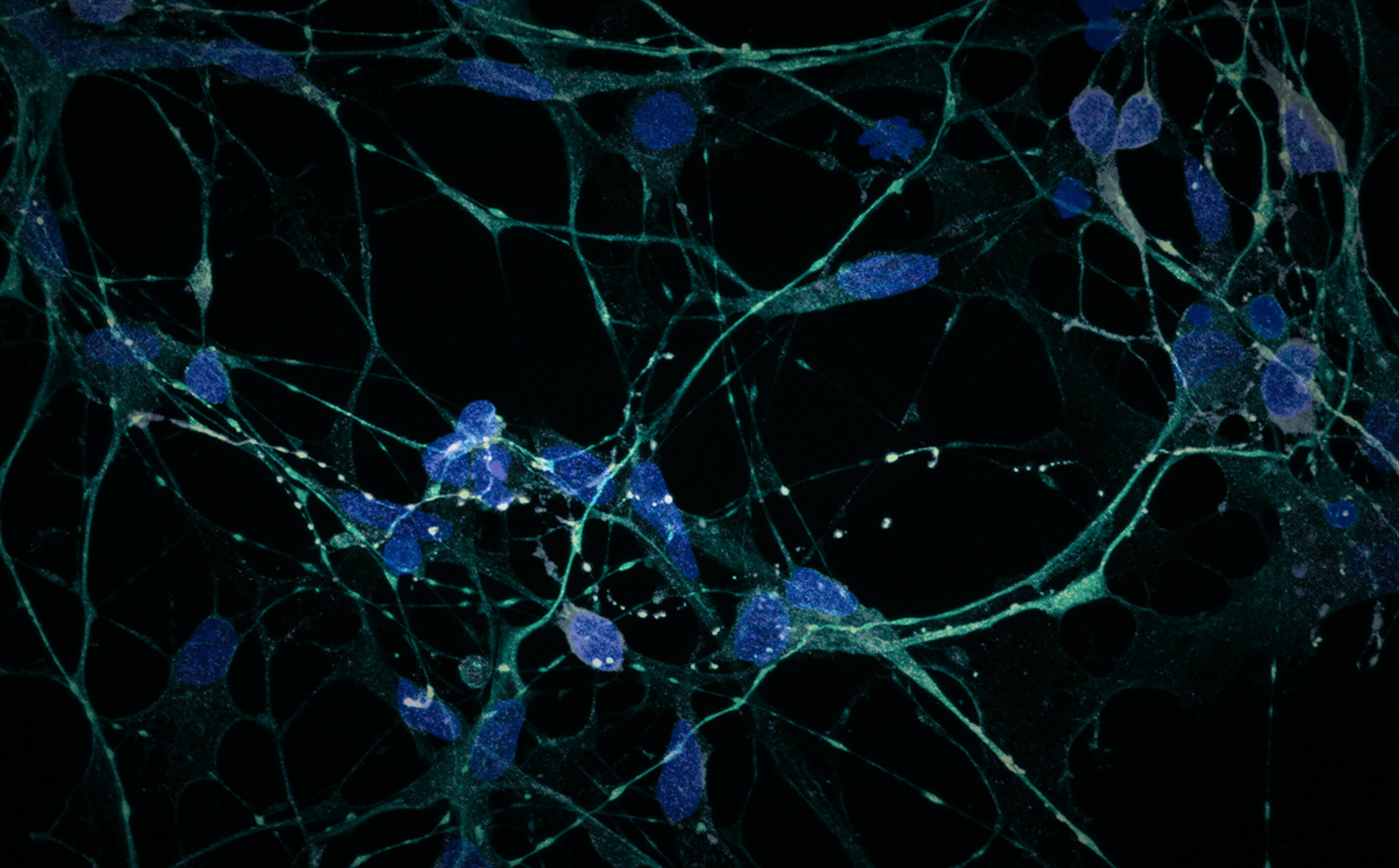
The Morphology and Imaging Core is a centralized facility serving the histological, high-content, live-cell, and in vivo microscopic imaging needs of all researchers and projects at the Buck Institute as well as external users. In addition to microscopy, bioenergetic assays such as cell respirometry are provided. The Core provides space, equipment, service, training and expert advice on many aspects of light and transmission electron microscopy from sample preparation through imaging to data analysis.
-
 Akos Gerencser, MD, PhD . Director
Akos Gerencser received his MD and PhD in neurosciences at Semmelweis University and his MS in biomedical engineering at Budapest University of Technology and Economics in Hungary. His postdoctoral work was at the Burnham Institute for Medical Research in La Jolla, California, and at the Buck Institute. He has worked with Drs. David Nicholls and Martin Brand at the Buck Institute since 2007 and since 2012 as an assistant research professor. His interdisciplinary research merges assay technology development (primarily advanced live-cell fluorescence microscopy) for mitochondrial physiology and bioenergetics with application of these technologies to neurodegenerative diseases, stem cell biology, and type 2 diabetes. He has developed the oxygen consumption rate calibration algorithm for the Seahorse Extracellular Flux Analyzer and a novel technology for unbiased, millivolts determination of mitochondrial membrane potential.
-
 Hao Cheng . Bioinformatics Scientist
Hao Cheng is a Bioinformatics Scientist with a master's degree in computer science. Hao develops skills in large-scale genomics data processing and analysis, involving statistical modeling, data visualization, machine learning, and in-house bioinformatics pipeline optimization. In addition, he had experience in deep learning and multi-omics analysis.
HCheng@buckinstitute.org
-
 Stella Breslin, BS . Senior Research Associate - Morphology
Stella Breslin graduated from Cal State East Bay in 2013 and joined the Buck Institute in 2014. She received a certificate in optical microscopy and advanced bioscience microscopy from the Merritt College Microscopy Program in 2010. She oversees and assists with all core responsibilities, including tissue processing and embedding and histological sectioning of frozen and paraffin samples. She is also involved in core imaging projects and shares general project management duties with collaborators.
-
 Harris Ingle . Research Associate
Harris Ingle graduated from UC Berkeley in 1994. He received a certificate in optical microscopy and advanced bioscience microscopy from Merritt College in 2010, where he continued to work as a lab technician for the Merritt Microscopy Program. He was in the first cohort of Merritt College’s Histotechnology program in 2015. He joined the Buck Institute in 2019 and assists with core services such as tissue embedding, sectioning, and histological staining. His duties include training the Institute’s technical and research staff in the use of facility instrumentation and software.
-
 Chad A. Lerner, PhD . Postdoctoral Researcher
Chad Lerner received his PhD in molecular biology at the Drexel College of Medicine in 2013, and completed a T32 NIH training fellowship at the University of Rochester aimed at exploring the effect of electronic cigarette oxidants on a critical animal lung antioxidant defense system and in mitochondria. His earlier graduate studies revealed mitochondrial involvement in the conferred benefit of converging pathways between rapamycin and low IFG-1 signaling on lifespan. His duties include training the Institute’s technical and research staff for cell respirometry and advanced live-cell and high-content microscopy and to carry out such assays as service.
CLerner@buckinstitute.org
-
 Shona Mookerjee, PhD . Adjunct Faculty and Research Scientist
Associate Professor, Touro University California College of Osteopathic MedicineShona Mookerjee has a scientific background is in molecular biology, genetics, and biochemistry, with a focus on mitochondrial biology. Her research program is currently focused on mitochondrial activity and energy transduction in different biological settings. She has more than 10 years of experience in experimental design and analysis using the Seahorse XF Analyzer with multiple biological models (including isolated mitochondria, cell culture, and whole organisms) in both academic and biotechnology settings. Her duties include advising and training on ATP flux measurements using the Seahorse XF Analyzers.
SMookerjee@buckinstitute.org
Laboratory services
- Tissue processing, embedding, and sectioning: frozen, paraffin, or floating
- Histological stains: H&E, Cresyl Violet, Oil Red O, etc.
- Antigen retrieval
- Immunohistochemistry or immunocytochemistry with enzyme-substrate development (HRP, AP) or fluorescent secondaries
Imaging services
Functional imaging:
- Calcium and other ion concentrations
- Fluorescent protein localization and aggregation
- Organelle motility
- Apoptosis, cell viability, migration
- Long-term time lapses
- FRET, FRAP
- Mitochondrial and plasma membrane potential, NADH
- Photoactivation/uncaging
High content imaging:
- Automated microplate-based multi-dimensional imaging
- Cell counting
- Cytometry
- Cellular senescence
Conventional microscopy:
- Brightfield, DIC, phase-contrast, fluorescence (blue/green/red/infrared)
Automated whole slide scanning (brightfield and fluorescence)
Laser scanning confocal and two-photon microscopy:
- Histological slides, microplates and deep-tissue; slice culture, fly organs, intact worms, intracranial imaging
- Immunolocalization, co-localization
- Fluorescence correlation spectroscopy
- Functional time course
Super resolution microscopy:
- Zeiss Airyscan2
- Super Resolution Radial Fluctuations
Transmission electron microscopy:
- Sample preparation, sectioning, imaging, image analysis
Image processing services:
- Zeiss Zen Desk: Measurement, Image Analysis, Extended Focus, 3Dxl, Intellesis
- Bitplane Imaris: modules include Imaris Core, Measurement Pro, FilamentTracer, co-localization and batch processing for 3D or 4D reconstruction and analysis for fixed or functional imaging data
- Image Analyst MKII: multidimensional time-lapse analysis, customized high content analysis, automated image analysis
Bioenergetics services:
- Mitochondrial isolation
- Seahorse Extracellular Flux Analysis for oxygen consumption and proton production rates and glycolytic and oxidative phosphorylation ATP fluxes
- Cell respirometry in suspensions
- Mitochondrial membrane potential assaying in cell cultures and tissue explants
- Mitochondria: cell volume fraction assay
- Mitochondrial ultrastructure with electron microscopy
- H2O2 production assay
Histology
Leica RM2155 embedding center, sliding, and automated rotary microtomes
Leica CM1950 cryostat
Microm HM505EP cryostat
RMC MT7000 ultramicrotome with diatome diamond knives
Leica TP1020 automated tissue processor
Light microscopy
Zeiss LSM 980 confocal microscope with 405 nm, 445 nm, 488 nm, 514 nm, 561 nm, 639 nm diode lasers, 32+2 channels Quasar spectral detector, PIEZO Z motorized stage, AI sample finder, Definite Focus 3, Airyscan2, heated environment chamber, CO2 control, additional Colibri7 light source and wide field camera. Equipped with constant temperature enclosure and CO2 regulation for functional assays.
Zeiss LSM 780 confocal with 405nm and 440nm Diode, Argon (458nm, 488nm and 514nm), 561 nm DPSS and 633 nm HeNe lasers for fixed or functional imaging. Equipped with constant temperature enclosure and CO2 regulation at the stage for functional assays.
Zeiss LSM 7MP deep tissue imager with Chameleon Vision II laser, (680nm-1080nm).
Zeiss LSM 700 on an upright AxioImager M2 with 405 nm, 488 nm, 555 nm, and 639 nm Diode lasers and motorized stage for fixed specimen imaging.
Nikon Eclipse Ti-PFS wide-field fluorescence microscope with TIRF and SRRF super resolution options, controlled by NIS Elements 5.20 or µManager and equipped with a Lambda 821 LED light source with Lambda 10-3 emission filter wheel an Andor iXon Life 888 EMCCD camera and an Air/CO2 gas mixer. Equipped with filter sets for NADH, Fura2, Fluo3, TMRM, CFP, GFP, YFP, RFP, Cy5, NIR750. Equipped for Phase contrast.
Nikon Eclipse Ti-PFS wide-field fluorescence microscope controlled by NIS Elements 5.20 and equipped with a Lambda LB-LS17 light source with Lambda 10-3 excitation and emission filter wheels a Cascade 512B EMCCD camera, a MS-2000 motorized stage and an OKOLab Air/N2/CO2 gas mixer. Equipped with filter sets for NADH, Fura2, Fluo3, TMRM, CFP, GFP, YFP, RFP, Cy5. Equipped for DIC.
Zeiss Axioscan 7 bright field and fluorescence whole slide scanner with 5x, 10x, 20x lenses, Colibri 7 LED light source (385 nm, 430 nm, 475 nm, 555 nm, 590 nm, 630 nm, 735 nm), Axiocam 705 color and Axiocam 712 monochrome cameras, 100 slides capacity.
Nikon Ni-E upright microscope with motorized stage, MQA18000 DS-Fi3 Microscope Camera controlled by NIS Elements 5.20, epifluorescence, DAPI, GFP, Texas Red and CY5 filters.
Nikon TE300 with DXM1200f CCD camera, DIC optics and Hg bulb for fixed sample brightfield, DIC and epifluorescence imaging.
Electron microscopy
Transmission electron microscopy: FEI-Philips Tecnai 12, tilting stage, Gatan Bioscan CCD
Bioenergetics and other
- Agilent Seahorse XFe24:24-well extracellular flux analyzer
- Agilent Seahorse XFe96:96-well extracellular flux analyzer
- Instech two-chamber oxygraph with phosphorescent O2 sensor for suspension cell and mitochondrial respirometry
- BMG Pherastar FS absorbance and fluorescence plate reader for H2O2 production assays
- Perkin Elmer EnSpire Alpha plate reader: luminescence and AlphaLISA/AlphaScreen microplate reader
We charge researchers at an hourly rate for utilizing core services and equipment.
External contracts are evaluated on a per-project basis.
Please contact the core manager for further information.
Contact:
Stella Breslin
Senior Research Associate – Morphology
SBreslin@buckinstitute.org
415-209-2000 ext 6809
Stella Breslin
Senior Research Associate
SBreslin@buckinstitute.org
415-209-2000 ext 6809
CORE TECHNOLOGIES
Technological innovation in the service of scientific advancement
Our cutting-edge technologies support the Institute’s goals and put the newest capabilities in the
hands of our scientists.