
by Buck Institute
November 18, 2026 . News
The hypothalamus is not a bystander in Alzheimer’s disease
Age-related changes in the brain’s body clock are linked to early symptoms of the disease, providing new targets for research and potential therapeutics
Alzheimer's disease is well known as a disease of memory. But long before memory fades, many patients have trouble sleeping, lose their daily rhythms, and show changes in metabolism and stress hormones. All of these functions are regulated by the hypothalamus, a structure at the base of the brain. A new study from the Buck Institute, published on November 18, 2026 in Nature, shows that the hypothalamus is not a bystander in Alzheimer's. Its circadian and metabolic control centers show specific age-related changes in volume and gene activity early in the disease.
 "Most of what we know about Alzheimer's comes from the hippocampus and cortex, because that's where memory is organized and lives," said senior author Ashley Webb, PhD, associate professor at the Buck. "But patients and families often report disrupted sleep, a change in appetite, or different responses to stress alongside memory changes.” Noting that recent studies even suggest that changes such as circadian disruptions may precede the development of other common clinical symptoms, Webb added, “We wanted to look directly at the part of the brain that controls those functions, and to do it with enough resolution to see which cells are changing with age and in the disease.”
The hypothalamus
The hypothalamus plays a vital role in maintaining physiological balance; it regulates various bodily functions such as temperature, sleep rhythms, hunger, thirst, and hormone release. The almond-shaped structure located above the roof of the mouth is made up of many specialized sub-areas called nuclei. Each nucleus runs a different job: the suprachiasmatic nucleus (SCN) is the body's master clock, the arcuate nucleus handles appetite and energy balance, and so on. Very little is known about how these individual nuclei, and their cell types change in human aging and Alzheimer’s. Webb’s team set out to fill that gap.
What the researchers did
The team built a resource called HypoAD, which is available to the research community, by combining two very different kinds of measurement:
- Brain imaging Using MRI scans from 458 living adults and a high-resolution atlas, the researchers measured the volume of 13 separate hypothalamic sub-regions. Participants were grouped by cognitive status and by levels of amyloid-β, the protein that forms plaques in Alzheimer's.
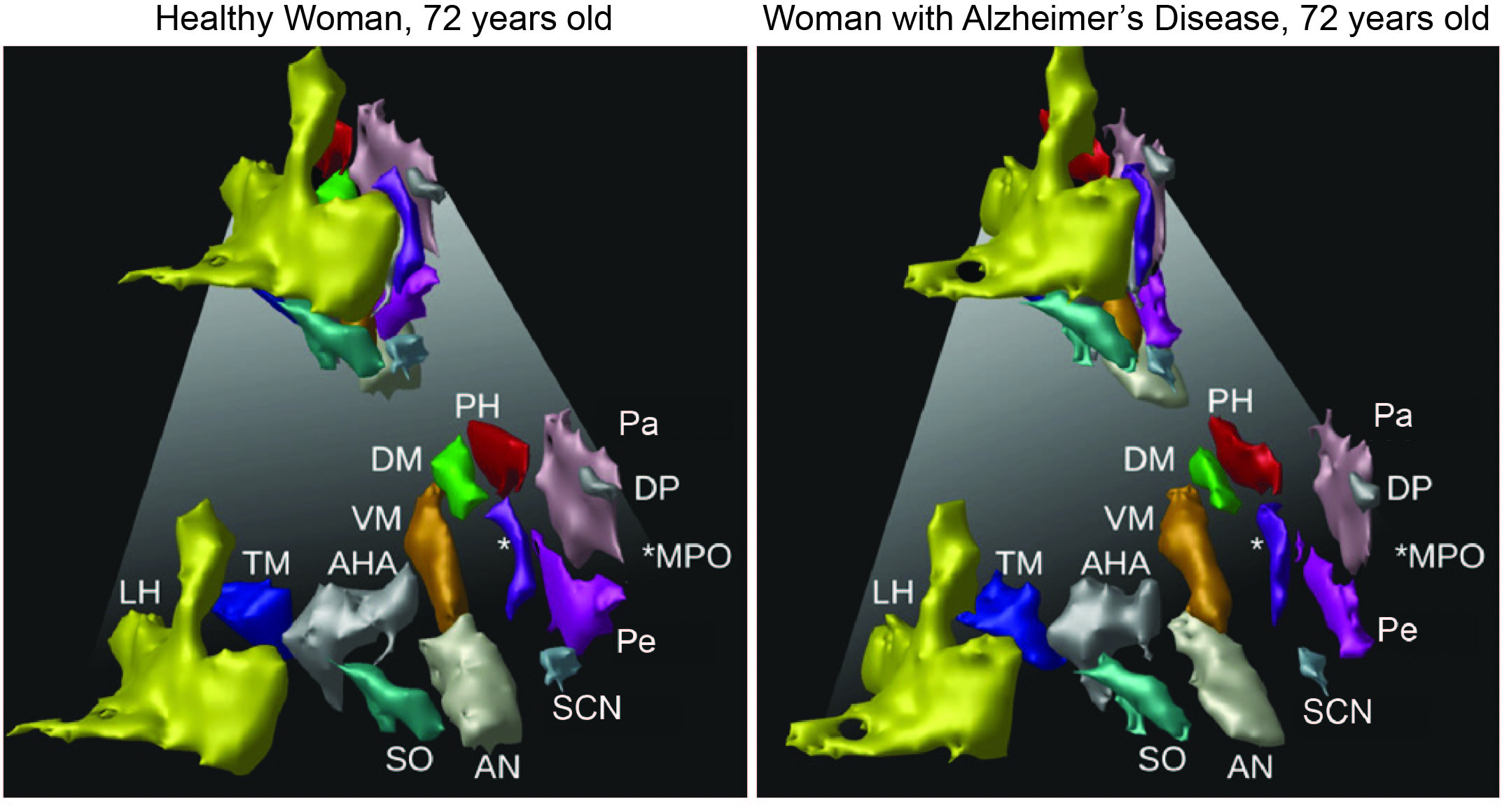
Three-dimensional reconstructions of hypothalamic subregions from MRI scans of two female individuals at 72 years of age, one healthy (left) and one with Alzheimer’s Disease (right). Differences in volume can be observed in regions involved in regions labeled SCN, AN, and Pe, which are important for sleep, circadian rhythms, and metabolic regulation.
Credit: Jürgen Germann
- Single-cell gene activity. The team sequenced about 614,000 individual cells from post-mortem hypothalamus tissue. Donors included seven people with mostly early-stage Alzheimer's, seven age-matched healthy controls, and three younger people from published data. This method shows which genes are switched on in each cell, so changes can be traced to specific cell types and cellular functions.
The researchers validated their findings against spinal fluid protein measurements, a prefrontal cortex dataset from about 800 donors, spatial gene-expression maps, and a mouse model of Alzheimer's.
 "No single method can answer this question," said co-senior author Manu Goyal, MD, director of neuroimaging labs at Washington University School of Medicine in St. Louis, MO. "MRI can tell you what's happening in living people but is limited in its resolution. Single-cell sequencing tells you what's happening inside each cell type but requires actual tissue. The goal here was to put them side by side to let us see things that neither could show alone."
Key Findings
Hypothalamic sub-regions age differently
The scientists found that though overall hypothalamic volume barely changed with age, different areas of the hypothalamus changed in opposite directions. The lateral hypothalamus shrank with age, while the SCN, arcuate, paraventricular, periventricular and dorsomedial nuclei grew. Webb says this phenomenon held after accounting for Alzheimer's status, suggesting it is a feature of typical aging. Volume differences tied specifically to AD also occurred but weren’t statistically significant, suggesting that a more sensitive method might be needed to determine what happens with the Alzheimer’s hypothalamus.
"If you only measure the hypothalamus as a whole, it looks like nothing is happening," Webb said. "When you zoom in, you see some regions shrinking and others growing. That tells us aging is reshaping this region in a very specific, structured way."
Single cell resolution gene expression measurements pick up what MRI misses.
Machine-learning models could tell Alzheimer's cells from control cells using gene expression alone, and the most important genes centered on two themes: inflammation and circadian rhythm.
Immune cells and clock neurons lead the changes
Microglia, the brain's resident immune cells, showed the largest number of altered genes. When the researchers ordered cells along a young-to-old-to-Alzheimer's path, microglia gradually shifted from a resting state toward the "disease-associated" state seen in other Alzheimer's studies. “This finding suggests that the hypothalamus transitions to an inflammatory state early in Alzheimer’s disease, just like the brain regions involved in memory,” said Webb.
The hypothalamus is a diverse area of the brain, harboring many distinct cell types critical for its varied physiological roles. The scientists identified 254 neuron subtypes and found that inhibitory (GABAergic) neurons changed the most, mainly in neuropeptide and receptor signaling. One prominent group of neurons in the SCN showed disrupted signaling by vasopressin, a key output of the body's clock.
"Sleep and circadian problems are some of the earliest signs of Alzheimer's, and circadian behavioral changes known as sundowning, occur later in the disease. But we've never had a good molecular explanation for why," Webb said. "Seeing vasopressin signaling disrupted in the brain's master clock, in people at early stages of the disease, gives us a concrete place to start."
There’s a possible reason some hypothalamic regions might grow
Total cell numbers did not appear to change. Instead, the data point to cells getting larger through increased activity of mTOR, a growth pathway, along with remodeling of the material between cells and inflammatory activation of glia. The authors present this as a hypothesis for future work.
What comes next
The authors note several limits to the work. The autopsy cohort is small, at 14 donors, though typical for this kind of study. The MRI and sequencing data come from different people, so the two are compared side by side rather than combined statistically. And because the design is cross-sectional, the results show associations, not cause and effect.
"This is a starting point, not the final word," Webb said. "Our next step is to follow these changes over time and test whether protecting the brain's clock, and neuropeptide signaling in particular, can slow the disease or help manage symptoms. If circadian disruption is one of the first things to go wrong, it may also be one of the first places we can intervene."
Citation
Hypothalamic volume and cell specific changes in aging and Alzheimer's disease.
DOI: 10.1038/s41586-026-11305-w
Additional collaborators:
Manu S. Goyal, Co-Senior Author, Department of Neurology, Washington University School of Medicine, St. Louis, MO: Doudou Yu, Buck Institute for Research on Aging; Ghulam Murtaza,, Michael J. MacCoss and William Stafford Noble, Department of Genome Sciences, University of Washington, Seattle, WA; Jürgen Germann, Institute of Biomedical Engineering, University of Toronto, Toronto, ON, Canada; Kaitlyn H. Hajdarovic ,Kelsey R. Babcock and Habil Zare, Neuroscience Graduate Program, Brown University, Providence, RI; Shiva Kazempour Dehkordi, Glenn Biggs Institute for Alzheimer’s and Neurodegenerative Diseases, San Antonio, TX, Ritambhara Singh, Department of Computer Science, Brown University, Providence, RI; Miranda E. Orr, Department of Neurology, Washington University School of Medicine, St. Louis, MO, Ivana Delalle, Department of Pathology and Laboratory Medicine, Boston University School of Medicine, Boston, MA: Alexander C. Jackson, Department of Physiology and Neurobiology, University of Connecticut, Storrs, CT; Ivana Delalle, Department of Pathology and Laboratory Medicine, Boston University School of Medicine, Boston, MA; Andrei G. Vlassenko, Mallinckrodt Institute of Radiology, Washington University School of Medicine, St. Louis, MO.
Acknowledgments
The work was supported by grants from the NIH/NIA: AG053503, AG057536, RF1 AG073210, R21 AG070527, AG078299, AG084624, R01 MH112739. AG078299, R01 AG068293, R01 AG068293, AG084624 and NIH/NIA F99/K00 award AG083292. Additional support came from the National Science Foundation, DBI 2213824. Support also came from the Blavatnik Family Graduate Fellowship in Biology and Medicine, the Carney Institute for Brain Science at Brown University, the Cure Alzheimer’s Fund, and the American Federation for Aging Research.
Science is showing that while chronological aging is inevitable, biological aging is malleable. There's a part of it that you can fight, and we are getting closer and closer to winning that fight.
Eric Verdin, MD, Buck Institute President and CEO